
How does the keratinous beak of birds affect their functional performance?
Bird beaks are a composite structure where a bony core is surrounded by a keratinous sheath, the rhamphotheca. It is the rhamphotheca that comes into direct contact with the birds’ environment, be that during feeding, preening, nest building, or any of the other numerous behaviours that are carried out using the beak; however, most morphological study that has been carried out on bird beaks has been conducted on the bony core only[1]. This is because in most skeletal museum preparations, the rhamphotheca becomes separated from the bone, and therefore a greater sample size can be obtained when the rhamphotheca is not included. If the shape of the rhamphotheca perfectly tracked that of the bony core, this wouldn’t be a problem, but we know that this is not the case. The rhamphotheca may extend greatly beyond the tip of the bony core, dramatically altering both the length and curvature of the beak overall. Moreover, we know that the covariation in shape between these two tissues differs between bird families: the prominent hooked beaks of raptors may have little bone extending into the beak tip, whereas flatter-beaked birds like kingfishers have a rhamphotheca that closely matches the shape of the underlying bone. Other birds may have tooth-like projections along the lateral edges of the keratin, or have large, hollow casques. Excluding these shape variations may have significant implications for how the functional morphology of the beak has been interpreted.
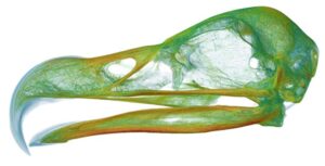
This project will use CT scans to reconstruct the bone and rhamphotheca of a number of bird beaks. The objectives are then to: 1) using geometric morphometrics[2], quantify the covariation between the bony and keratinous beak across a wide range of bird taxa, establishing how this relationship varies among families; 2) use this knowledge to explore the functional consequences of rhamphotheca changes in shape. This latter question will be explored via hypothetical deductive finite element analysis (FEA)[3], where variables such as the thickness and curvature of the rhamphotheca will be adjusted in different areas to explore how this affects beak strains. We already know that the presence of a rhamphotheca acts to reduce skull strains overall[4], but a comprehensive assessment of how specific morphological changes affect the patterns and magnitudes of strain in the beak is lacking. This has implications for the modelling of all beaked taxa, but is particularly useful for understanding extinct beaked taxa (e.g. dinosaurs) where the precise soft-tissue morphology can be poorly known. With a better understanding of how the hard and soft tissues of the beak evolve together, we will be able to gain a greater appreciation for the biodiversity present in this structure, and in the multiple reptilian taxa in which it has evolved through time.
This project is part of a larger multidisciplinary study investigating functional performance and its evolution in the feeding mechanics of bird beaks and skulls. The student will work with a team spanning Biology and Engineering, and will have access to the resources of both, including a high-performance computer cluster, materials testing facilities, and a micro-CT scanner. The student will receive training in the digital reconstruction of anatomy from CT and diceCT[5] scans, and how to use these reconstructions in downstream finite element modelling. References: [1] Bright et al. 2016. PNAS. 113:5352-5357; [2] Adams et al. 2013. Hystrix. It. J. Mamm. 24:7-14; [3] Rayfield. 2007. Ann Rev Earth Planet Sci 35:541-576; [4] Lautenschlager et al. 2013. PNAS. 110:20657-20662; [5] Lautenschlager et al. 2014. J Anat 224:412-431